Printed from acutecaretesting.org
June 2002
Key elements in a successful pediatric laboratory
Introduction
Children, especially infants, are not simply small people. A laboratory that serves children must understand this. The patient population in pediatrics is extremely diverse, ranging from premature babies and other neonates to children in all stages of development, including adolescents.
The challenges facing the pediatric laboratory range from the collection of specimens, and the necessity of using very small volumes of serum or plasma, to differences in reference ranges.
Another important facet of the pediatric laboratory is the facility to test for metabolic and genetic diseases.
In addition, children’s conditions are often more labile than those of adults, which creates the need for rapid testing, often at the bedside (point-of-care testing).
Adolescents, even when seemingly healthy, are subject to many social diseases or conditions. The use of drugs of abuse is most common in this age group. Teenage pregnancy, sexually transmitted diseases, and suicide are all factors that have an impact on the pediatric laboratory.
This paper will focus primarily on the needs of neonates and infants in Pediatric Intensive Care Units (PICU).
The newborn is highly vulnerable to the physiological changes needed for extrauterine life. This is particularly true for the premature infant who enters life too early in the gestational period. The highest risk neonates are those born at less than 1 kg at birth. In the Neonatal Intensive Care Unit (NICU) at our hospital, we see about 90-100 such infants yearly. Our NICU has 40 beds, which are almost always in use.
The major conditions seen are prematurity, hyaline membrane disease, sepsis, pneumonia, meconium aspiration, and congenital heart disease. Though less common, metabolic and other genetic diseases create a challenge to the neonatologist.
In recent years, the introduction of Extracorporeal Membrane Oxygenation (ECMO) has saved the lives of many prematures presenting with meconium aspiration, as well as for those neonates with postsurgical cardiac failure. ECMO essentially effects cardiac support for those children who have reversible respiratory failure.
In our PICU, which has 16 beds, the most common conditions seen are also related to pulmonary or cardiac diseases. Those most frequently seen include: respiratory dysfunction, postoperative cardiopulmonary or other postoperative conditions, pneumonia, bronchiolitis, metabolic disturbances, and sepsis.
Blood collection
Blood drawing is a major challenge in pediatric medicine, especially from premature infants and neonates.
Skin puncture is the preferred method for neonates and small infants. Heel sticks are usually used for premature babies and other neonates, and finger sticks for infants and young children.
 |
FIG. 1: Sites of Collection from an Infant’s Heel.
From: Blumenfeld TA, Tri GK, Blanc WA. Recommended site and depth of newborn heel skin punctures based on anatomical measurements and histopathology. Reprinted with permission from Elsevier Science (The Lancet 1979; 1: 231).
In 1986, the National Committee for Clinical Laboratory Standards (NCCLS) published “Procedures for the Collection of Diagnostic Blood Specimens by Skin Puncture” [1]. The sites of collection from an infant’s heel were recommended by Blumenfeld [2], and are reproduced from the book by Soldin, Rifai and Hicks [3]. The exact details, as outlined in these references, are illustrated graphically in Fig. 1.
Venipuncture is the method of choice for the collection of blood from older children, who are less likely to be traumatized by the sight of a needle than younger children! Specimen volume is of great concern for premature infants and children.
As mentioned above, we take care of many babies under 1 kg in weight, whose total blood volume is less than 115 mL. Hicks [4] recently published a letter in the New England Journal of Medicine describing her survey of the amounts of blood drawn for various tests in major teaching and community hospitals.
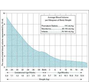
Her conclusion was that many laboratories draw far more blood than is necessary, and this practice is unacceptable for pediatric patients. Fig. 2 shows the average blood volume by age for prematures, newborns, and infants. Table I shows the hematocrit by age.
It is obvious that, as hematocrits are high and blood volumes low, it is essential to keep blood drawing to a minimum, otherwise replacement transfusions may be necessary.
 |
|
FIG. 2: Infant Blood Volume.
From Werner M, ed. Microtechniques for the clinical laboratory. New York: Wiley, 1976:2. Reprinted with permission.
|
TABLE I: Hematocrit By Age. From Matoth Y. Zaiaov R, Varsaro I. Acta Paed. Scand 1971;60:318. Reproduced with permission. |
The other major consideration with small specimens is the effect of leaving such samples uncovered, which can lead to evaporation of the liquid in the sample [5]. For example, it is generally observed that evaporation can easily lead to a 10 % fluid decrease from a 100 µL specimen.
The choice of instrumentation is critical, although much easier now than 20 years ago. It is preferable to have all analyses performed on 20 µL or less of serum or plasma. It is also important to understand the significance of the “dead volume” required in the sample cup of automated analyzers, that is, the amount of sample that remains in the cup after samples are aspirated for analyses.
Obviously, these considerations are less serious in laboratories devoted to adult patients.
Metabolic and genetic diseases
This topic requires a full paper in itself, so it will be discussed only briefly here.It is important to note that it is in the neonatal period that many of these diseases become manifest, and if not treated expeditiously, can result in mental retardation or death. Some such diseases include: galactosemia, maple syrup urine disease and ornithine transcarbamoylase, to mention only a few.
A metabolic disease should always be considered in a neonate with “failure to thrive” and unexplained metabolic acidosis. Hepatosplenomegaly, muscle weakness and bone abnormalities may also lead one to suspect a metabolic disease. The first line of testing includes analyses of amino acids and organic acids.
With the recent elucidation of the human genome, it is clear that many more gene-linked diseases will be identified, and that the methods of choice for diagnosis of such diseases will consist of molecular diagnostic techniques, such as the use of DNA chips and single nucleotide polymorphisims (SNP).
Reference ranges
Again, this is a topic for an entire paper, but suffice it to say here that many test values change dramatically with age. For example, the reticulocyte count is 2.2-4.8 % from 1-3 days after birth to 0.4-2.7 % at 4-30 days after birth. Thyroxine is 3.0-14.4 µg/dL at 1-30 days to 5.3-16.3 µg/dL at 1-20 months.
The reader is directed to reference [6] that is dedicated to this topic. It is obviously necessary to have age-related reference value ranges, otherwise unnecessary additional testing could be ordered.
Point-of-care testing (POCT)
This type of testing is particularly suited to testing in the neonatal and infant intensive care units. As many prematures, neonates, and infants have serious respiratory problems, it is critical to have available rapid blood gas and electrolyte measurements that can be performed at or near the patient’s bedside.
When neonates are on ECMO, they need frequent monitoring of pH, pCO2 and pO2, usually every 2 hours, depending on the case. The measurement of electrolytes is also essential to correct abnormalities of potassium and/or calcium, in order to avoid any possible arrhythmias prior to the neonate being placed on ECMO. As dehydration often occurs in prematures and other neonates, rapid measurements of sodium and potassium must be available.
There are several possible approaches to POCT for the above-described situations. An attractive is a hand-held device. Another approach is the use of a benchtop bloodgas analyzer. We use both instruments in different settings. In-line testing of blood gases is obviously an excellent approach for patients on ECMO.
Our neonatologists have evaluated such sensors, and they have demonstrated excellent correlation with our usual methodology [7]. It is almost certain that, in the future, we will be able to measure blood gases non-invasively. In our NICU and PICU, all of the blood gas measurements are performed by POCT instruments. In our NICU and PICU, 61 % and 93 %, respectively, of electrolytes are done by POCT.
The monitoring of blood gases is also important in the operating room, particularly in open-heart surgery. The results must be ready immediately if they are to be of any value to the surgical team.
Activated blood-clotting time measurements are often critical both for neonates on ECMO and children undergoing open-heart surgery. During ECMO, the test should be performed every 30-60 minutes, depending on the stability of the values or any bleeding complications. The tests must be performed immediately; otherwise values will not be accurate.
The use of POCT in the NICU and PICU extends far beyond blood gases and electrolytes. Glucose measurements are often of great importance, as hypoglycemia is encountered frequently in prematures and other neonates, particularly when hepatic function is not fully developed and, therefore, gluconeogenesis is compromised.
In addition, we have the approximately 900 diabetic children seen at our hospital who require not only glucose measurements but glycosylated hemoglobin and microalbumin values.
Many other tests can also be performed by POCT. These include, for example, tests such as Group A streptococcus, sexually transmitted diseases, pregnancy, drugs of abuse.
It can be seen that the key elements for a pediatric laboratory stem from an understanding that children are different from adults. The pediatric laboratory, therefore, must be planned and directed differently.
The future will bring many challenges as we embrace POCT for all basic testing and move to more exotic testing such as molecular diagnostics and tandem mass spectrometry. We must embrace change as we move through this new century.
References+ View more
- Procedure for the collection of diagnostic blood specimens by skin puncture. 2nd ed. NCCLS Approved Standards, 1986.
- Blumenfeld TA, Turi GK, Blanc WA. Recommended site and depth of newborn heel stick punctures based on anatomical measurements and histopathology. Lancet 1979, 1: 230-33.
- Hicks JM. In: Biochemical Basis of Pediatric Disease. Soldin SJ, Rifai N, Hicks JM, eds. Washington, DC: AACC Press: 1-16.
- Hicks JM. Excessive blood drawing for laboratory tests. N Eng J Med 1999; 340: 1690 (letter).
- Rifai N. Personal communication.
- Soldin SJ, Brugnana C, Hicks JM. Pediatric Reference Ranges. 3rd ed. Washington, DC: AACC Press, 1999.
- Bahrami K, Personal communication.
References
- Procedure for the collection of diagnostic blood specimens by skin puncture. 2nd ed. NCCLS Approved Standards, 1986.
- Blumenfeld TA, Turi GK, Blanc WA. Recommended site and depth of newborn heel stick punctures based on anatomical measurements and histopathology. Lancet 1979, 1: 230-33.
- Hicks JM. In: Biochemical Basis of Pediatric Disease. Soldin SJ, Rifai N, Hicks JM, eds. Washington, DC: AACC Press: 1-16.
- Hicks JM. Excessive blood drawing for laboratory tests. N Eng J Med 1999; 340: 1690 (letter).
- Rifai N. Personal communication.
- Soldin SJ, Brugnana C, Hicks JM. Pediatric Reference Ranges. 3rd ed. Washington, DC: AACC Press, 1999.
- Bahrami K, Personal communication.
May contain information that is not supported by performance and intended use claims of Radiometer's products. See also Legal info.
Acute care testing handbook
Get the acute care testing handbook
Your practical guide to critical parameters in acute care testing.
Download nowRelated webinar
Evolution of blood gas testing Part 1
Presented by Ellis Jacobs, PhD, Assoc. Professor of Pathology, NYU School of Medicine.
Watch the webinar


