Printed from acutecaretesting.org
October 2003
Pulse oximetry vs. transcutaneous monitoring in neonates: practical aspects


Introduction
Non-invasive monitoring of oxygenation has become a standard procedure. Of the two methods available, namely transcutaneous partial pressure of oxygen (tcpO2) monitoring and pulse oximetry, the former is currently at risk of being replaced by the latter.
This is despite the fact that both methods provide substantially different information about oxygenation and are particularly helpful if used in combination.
This article reviews the principles of both techniques and discusses methodological and practical issues related to the use of these devices in infants.
Transcutaneous partial pressure of oxygen (tcpO2) monitoring
Principle of operation
tcpO2 electrodes measure the partial pressure of oxygen through the skin. They consist of a platinum cathode and silver reference anode, encased in an electrolyte solution and separated from the skin by an O2-permeable membrane.
Electrodes are heated to improve oxygen diffusion and to arterialize the capillary blood. Oxygen is reduced at the cathode, generating an electric current proportional to the O2 concentration in the capillary bed underneath the sensor.
Sensors require a 10-15 min warm-up period after application and have to be calibrated every 4-8 hours.
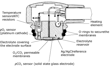
FIG 1. Structure of a combined tcpO2/pCO2 transcutaneous electrode.
Factors influencing measurements
The agreement between arterial and skin-surface pO2 depends on a fragile balance between factors that increase the pO2, namely a shift to the right of the oxygen dissociation curve, and a decreased O2 solubility in blood, both of which are caused by the heating of the skin, and factors which decrease pO2, namely the oxygen consumption in the heated skin and inside the electrode [1].
Thus, transcutaneous (tcpO2) and arterial (pO2(aB)) represent two different measurements, and at times changes in tcpO2 are more important than the actual values measured. In hemodynamically unstable children, tcpO2 will reflect changes in the circulatory status and thereby give an early warning of the onset of circulatory compromise.
Sensor temperature
There is good agreement with arterial pO2 (pO2(aB)) only at 44 °C, but then frequent (every 2-4 hours) resiting is necessary. At lower sensor temperatures, tcpO2 will underread pO2(aB), with the difference becoming larger with increasing pO2(aB).
This is particularly important in preterm neonates, in whom high pO2(aB) levels must be reliably detected to minimize the risk of retinopathy of prematurity.
Probe placement
tcpO2 will underread pO2(aB) if the sensor is placed on a bony surface, if pressure is applied on the sensor, or if too much contact gel is used. With patent ductus arteriosus and right-to-left shunt, tcpO2 will be higher on the upper than on the lower half of the thorax.
In infants with a closed arterial duct, the electrode should ideally be placed on the lower back or abdomen, or on a thigh.
Peripheral perfusion
tcpO2 depends on skin perfusion. If the latter is reduced, e.g. due to hypotension, anemia, acidosis (pH < 7.05), hypothermia, or marked skin edema, tcpO2 will be accordingly low.
If an underreading of pO2(aB) occurs, it is advisable to check the patient for these conditions [2].
Skin thickness
Close agreement with pO2(aB) can only be found in neonates. This does not imply, however, that tcpO2 monitors can only be used in this age group.
Studies in children and adults did in fact show that the ratio between tcpO2 and pO2(aB) is extremely constant in these patients (independent of age and pO2(aB)), it merely is 20 % lower than in neonates, i.e. approximately 0.8 [3].
Detection of hypoxemia and hyperoxemia
Under optimal measurement conditions, tcpO2 can be expected to be within ±9.8-15 mmHg/1.3-2.0 kPa of pO2(aB) 95 % of the time [4]. Clinically, however, it seems more important to know whether the tcpO2 monitor will reliably detect all situations where a patient has either too little (e.g. < 40 mmHg/5.3 kPa) or too much oxygen (> 80 mmHg/10.7 kPa).
In this regard, the data available suggest that approximately 15 % of both hypoxemic and hyperoxemic instances are missed by tcpO2 monitors, whereas their specificity, particularly with regard to hypoxemia, is somewhat higher [5].
Pulse oximetry (sO2(p))
Principle of operation
Pulse oximeters do not measure the concentration of oxygen dissolved in plasma but the proportion of hemoglobin molecules in arterial blood that are loaded with oxygen. Deoxygenated hemoglobin absorbs more light in the red band (at 600-750 nm), i.e. it looks less red, whereas oxygenated hemoglobin absorbs more light in the infrared band (850-1000 nm).
The ratio of the absorbance of red and infrared light sent through a tissue correlates with the proportion of oxygenated to deoxygenated hemoglobin in the tissue.
Conventional pulse oximeters determine the arterial component within this absorbance by identifying the peaks and troughs in the absorbance over time, thereby obtaining a "pulse-added" absorbance that is independent of the absorbance characteristics of the non-pulsating parts of the tissue.
These pulse-added light absorbances are then associated algorithmically with empirically determined arterial oxygen saturation (sO2(aB)) values [1].
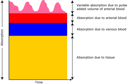
FIG 2. Conventional pulse oximetry : Light passes through the layers of the measuring site.
When the light absorption at the trough (which should include all the constant absorbers) is subtracted from the light absorption at the peak then, in theory, the resultants are the absorption characteristics due to added volume of arterial blood only.
Next-generation instruments use additional and/or different techniques.
For example, one recently developed technology scans through all red-to-infrared ratios (and corresponding sO2(p) values) found in the tissue, determines the intensity of these and chooses the right-most peak of these intensities, which will correspond to the absorbance by the arterial blood in the tissue.
It also uses frequency analysis, time domain analysis and adaptive filtering to establish a 'noise reference' in the detected physiological signal [6], thereby improving the ability to separate between signal and noise (see below).
Other next-generation instruments use differential signal amplification to achieve this goal.
Factors influencing measurements
Pulse oximeters are easier to use than tcpO2 monitors: they do not require calibration or heating of the skin and provide immediate information about arterial oxygenation.
However, it is probably because of this apparent ease of use that potentially erroneous measurements on a pulse oximeter are more at risk of being overlooked than those occurring with a tcpO2monitor.
A thorough understanding of the factors potentially affecting the precision of a pulse oximeter is therefore particularly important [1].
Probe placement
The light-receiving diode must be placed exactly opposite the emitting diode, and both must be shielded against ambient light and not be applied with too much pressure.
Light bypassing the tissue can cause both falsely high and falsely low values. The sensor site must be checked every 6-8 hours. It was recently shown that 42 % of nurses in a neonatal intermediate-care nursery exceeded a pressure on the skin of 50 mmHg/6.7 kPa during fixation of a pulse oximeter sensor [7].
Such high pressures may result in a reduced signal-to-noise ratio and may thus severely impair the precision of the sO2(p) measurements [7]. Highly flexible sensors provide better skin contact and thus better signal-to-noise ratio.
Peripheral perfusion
Conventional oximeters require a pulse pressure above 20 mmHg/2.7 kPa or a systolic blood pressure above 30 mmHg/4 kPa to operate reliably [8].
Because next-generation oximeters rely less on pulse detection, they continue to operate even at lower blood pressure levels [9].
Response times
In theory, the response time of a pulse oximeter depends only on the time it takes for the blood to travel from the lung to the sensor site, which is 3-4 sec in neonates [1].
However, all pulse oximeters currently available average their values over 2-15 sec to level out any erroneous measurement which may occasionally occur even under optimal conditions. This averaging, however, has unwanted consequences:
- it delays the response to a true fall in sO2(p) values
- it may lead to a mixing up of true with falsely low sO2(p) readings during periods of intermittent body movements (e.g. during feeding), which can result in the erroneous impression that the patient suffers episodes of prolonged hypoxemia
- it can lead to erroneous conclusions in situations where a precise measurement of sO2(p) is required, e.g. during sleep studies
- it hinders the definition of normal ranges for the frequency and severity of intermittent falls in sO2(p) in infants or children, since such data would only be valid for the specific averaging mode with which they have been obtained. This is particularly true for the next-generation instruments, which use variable averaging times depending on measurement conditions, i.e. 2-4 sec averaging under optimal conditions, and up to 15 sec averaging during periods of motion.
Motion artifact
The pulsatile (=arterial) component contributes less than 1 % to the total absorbance measured by the pulse oximeter.
Hence, at least conventional pulse oximetry is very sensitive to sudden changes in background signal, e.g. due to body movements.
Next-generation instruments use various techniques to identify and read through periods with low signal-to-noise ratios as there are during motion, resulting in dramatic (> 90 %) decreases in false alarm rates [10,11].
However, some of these improvements are achieved at the expense of not identifying true desaturation [12], which is unacceptable.
With conventional oximeters, identification of motion artifact is best done by displaying the light plethysmographic waveforms. Whenever these are distorted, sO2(p) readings become unreliable.
Alternatively, the pulse rate can be compared with the heart rate from an ECG monitor, which should be identical [13].
For next-generation instruments, which are less reliant on a clean peak-and-trough detection and thus an undisturbed pulse waveform, there is currently no independently validated method to identify periods of poor measurement conditions and thus potentially unreliable sO2(p) readings.
An interesting approach in this regard is the signal quality indicator, recently shown reliably to exclude erroneous measurements if above a certain threshold [14].
Other hemoglobins and pigments
Methemoglobin (MetHb) will cause sO2(p) readings to tend towards 85 %, independent of sO2(aB). Carboxyhemoglobin (COHb) will cause overestimation of sO2(aB) by 1 % for each percent COHb in the blood.
Fetal hemoglobin (HbF) and bilirubin do not affect pulse oximeters but may lead to a bias on sO2(aB) by CO-oximeters. In patients with dark skin, sO2(p) values may be falsely high, particularly during hypoxemia [1].
Algorithms
Pulse oximeters, in contrast to CO-oximeters, do not measure O2 saturation but derive their values from a "look-up" table which is based on empirical data from healthy adults.
These may vary between brands and even between different software versions from the same manufacturer. Also, some instruments subtract a priori the typical levels of COHb, MetHb, etc. in healthy non-smoking adults from their measurements and will thus display sO2(p) values that are some 2-3 % lower than those displayed by other instruments.
This approach, i.e. to display the so-called "fractional" sO2(p) instead of the usual "functional" sO2(p), has largely been abandoned in recent years, probably because it resulted in an unacceptably poor ability of instruments using this approach to detect hyperoxemia [15].
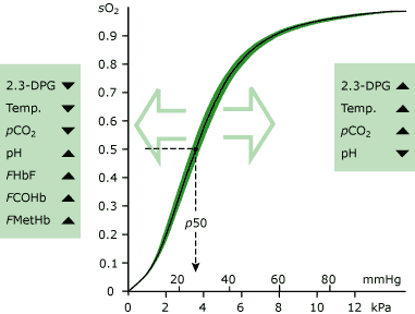
FIG. 3. The Oxygen Dissociation Curve illustrates the relationship between sO2(aB) and pO2(aB). Various conditions can shift the curve left or right as described in the boxes.
Detection of hypoxemia and hyperoxemia
In the absence of motion, pulse oximeters have both a high sensitivity and specificity for the detection of hypoxemia (sO2(aB) <80 %), although they tend to overestimate sO2(aB) during extreme hypoxemia (sO2(aB) <70 %) [1].
Due to the shape of the O2 dissociation curve, however, they are less well suited for detecting hyperoxemia. The upper alarm limits that have to be chosen on individual instrument brands to avoid hyperoxemia reliably range from 88 to 95 % [1,15].
An upper alarm limit of 95 % was recently confirmed for three next-generation instruments [16].
The reliability in detecting sO2(p) values >80 mmHg/10.7 kPa via non-invasive monitoring can likely be increased if both sO2(p) and tcpO2 are monitored.
tcpO2 or pulse oximetry?
Pulse oximeters and tcpO2 monitors measure different, but related variables. The relationship between pO2(aB) and sO2(aB), expressed in the oxygen dissociation curve, is neither linear nor constant.
For example, at high sO2(aB) levels, large changes in pO2(aB) may occur with very small changes in sO2(aB). Also, at a constant pO2(aB) of, for example, 45 mmHg/6 kPa, sO2(aB) may be either 80 % or 88 %, depending on whether arterial pH is 7.25 to 7.40.
The actual relationship between sO2(aB) and pO2(aB) and/or between tcpO2 and sO2(p) should, therefore, be repeatedly determined, particularly in critically ill patients.
In addition, both tcpO2 monitors and pulse oximeters have their specific shortfalls: the tcpO2 monitor is relatively difficult to use, has frequent periods where there is no reliable measurement (during warm-up and calibration periods) and reacts relatively slowly to sudden changes in oxygenation, whereas at least conventional pulse oximeters fail to operate reliably whenever the patient moves, are relatively prone to erroneous measurements resulting from improper probe placement and may be unreliable under conditions of hyperoxemia and possibly also severe hypoxemia.
For these reasons, both devices should ideally be used in combination, particularly whenever a reliable detection of hyperoxemia is paramount, as it is in extremely low-birthweight infants receiving additional inspired oxygen. In these patients, it may be advisable to reduce the FIO2 whenever one of the two instruments alarms.
References+ View more
- Poets CF, Southall DP. Non-invasive oxygen monitoring in infants and children: practical considerations and areas of concern. Pediatrics 1994; 93: 737-46
- Versmold HT, Linderkamp O, Holzmann M, Strohhacker I, Riegel K. Transcutaneous monitoring of pO2 in newborn infants: Where are the limits? Influence of blood pressure, blood volume, blood flow, viscosity and acid base state. Birth defects: original article series, Vol. XV, No. 4. New York: Alan R. Liss, 1979: 285-94
- Vyas H, Helms P, Cheriyan G. Transcutaneous oxygen monitoring beyond the neonatal period. Crit Care Med 1988; 16: 844-47
- Martin RJ, Robertson SS, Hopple MM. Relationship between transcutaneous and arterial oxygen tension in sick neonates during mild hyperoxemia. Crit Care Med 1982; 10: 670-72
- Poets CF. Monitoring in the NICU. In: Mathew OP. Respiratory control and its disorders in the newborn. New York: Marcel Dekker, Inc, 2003: 217-35
- Barker, SJ, Shah NK. Effects of motion on the performance of pulse oximeters in volunteers. Anesthesiol 1997; 86: 101-08
- Bucher HU, Keel M, Wolf M, von Siebental K, Duc G. Artifactual pulse-oximetry estimation in neonates. Lancet 1994; 343: 1135-36
- Morris RW, Nairn M, Torda TA. A comparison of fifteen pulse oximeters. Part I: A clinical comparison; part II: A test of performance under conditions of poor perfusion. Anaesth Intens Care 1989; 17: 62-82
- ECRI. Evaluation: Next-generation pulse oximetry. Health Devices 2000; 29: 347-79
- Bohnhorst B, Poets CF. Major reduction in alarm frequency with a new pulse oximeter. Intensive Care Med 1998; 24: 277-78
- Rheineck-Leyssius AT, Kalkman CJ. Advanced pulse oximeter technology compared to simple averaging. II. Effect on frequency of alarms in the postanesthesia care unit. J Clin Anesth 1999; 11: 196-200
- Bohnhorst B, Peter CS, Poets CF. Pulse oximeters' reliability in detecting hypoxemia and bradycardia: Comparison between a conventional and two new generation oximeters. Crit Care Med 2000; 28: 1565-68
- Poets CF, Stebbens VA. Detection of movement artifact in recorded pulse oximeter saturation. Eur J Pediatr 1997; 156: 808-11
- Urschitz MS, von Einem V, Seyfang A, Poets CF. Use of pulse oximetry in automated O2 delivery to ventilated infants. Anesthesiol 2002; 94: 37-40
- Bucher HU, Fanconi S, Baeckert P, Duc G. Hyperoxemia in newborn infants: detection by pulse oximetry. Pediatrics 1989; 84: 226-30
- Bohnhorst B, Peter C, Poets CF. Detection of hyperoxaemia in neonates: data from 3 new pulse oximeters. Arch Dis Child Fetal Neonatal Ed 2002; 87: 217-19
References
- Poets CF, Southall DP. Non-invasive oxygen monitoring in infants and children: practical considerations and areas of concern. Pediatrics 1994; 93: 737-46
- Versmold HT, Linderkamp O, Holzmann M, Strohhacker I, Riegel K. Transcutaneous monitoring of pO2 in newborn infants: Where are the limits? Influence of blood pressure, blood volume, blood flow, viscosity and acid base state. Birth defects: original article series, Vol. XV, No. 4. New York: Alan R. Liss, 1979: 285-94
- Vyas H, Helms P, Cheriyan G. Transcutaneous oxygen monitoring beyond the neonatal period. Crit Care Med 1988; 16: 844-47
- Martin RJ, Robertson SS, Hopple MM. Relationship between transcutaneous and arterial oxygen tension in sick neonates during mild hyperoxemia. Crit Care Med 1982; 10: 670-72
- Poets CF. Monitoring in the NICU. In: Mathew OP. Respiratory control and its disorders in the newborn. New York: Marcel Dekker, Inc, 2003: 217-35
- Barker, SJ, Shah NK. Effects of motion on the performance of pulse oximeters in volunteers. Anesthesiol 1997; 86: 101-08
- Bucher HU, Keel M, Wolf M, von Siebental K, Duc G. Artifactual pulse-oximetry estimation in neonates. Lancet 1994; 343: 1135-36
- Morris RW, Nairn M, Torda TA. A comparison of fifteen pulse oximeters. Part I: A clinical comparison; part II: A test of performance under conditions of poor perfusion. Anaesth Intens Care 1989; 17: 62-82
- ECRI. Evaluation: Next-generation pulse oximetry. Health Devices 2000; 29: 347-79
- Bohnhorst B, Poets CF. Major reduction in alarm frequency with a new pulse oximeter. Intensive Care Med 1998; 24: 277-78
- Rheineck-Leyssius AT, Kalkman CJ. Advanced pulse oximeter technology compared to simple averaging. II. Effect on frequency of alarms in the postanesthesia care unit. J Clin Anesth 1999; 11: 196-200
- Bohnhorst B, Peter CS, Poets CF. Pulse oximeters' reliability in detecting hypoxemia and bradycardia: Comparison between a conventional and two new generation oximeters. Crit Care Med 2000; 28: 1565-68
- Poets CF, Stebbens VA. Detection of movement artifact in recorded pulse oximeter saturation. Eur J Pediatr 1997; 156: 808-11
- Urschitz MS, von Einem V, Seyfang A, Poets CF. Use of pulse oximetry in automated O2 delivery to ventilated infants. Anesthesiol 2002; 94: 37-40
- Bucher HU, Fanconi S, Baeckert P, Duc G. Hyperoxemia in newborn infants: detection by pulse oximetry. Pediatrics 1989; 84: 226-30
- Bohnhorst B, Peter C, Poets CF. Detection of hyperoxaemia in neonates: data from 3 new pulse oximeters. Arch Dis Child Fetal Neonatal Ed 2002; 87: 217-19
May contain information that is not supported by performance and intended use claims of Radiometer's products. See also Legal info.


Acute care testing handbook
Get the acute care testing handbook
Your practical guide to critical parameters in acute care testing.
Download now
Related webinar
Combining blood gas invasive and non-invasive monitoring for the best care of our neonates
Webinar presented by Dr. Kaare E. Lundstrøm, Pediatric emergency and intensive care unit, Rigshospitalet, Denmark Watch the webinarRelated webinar
TC monitoring in NICU - the value of tcpO2
Presented by Daniele De Luca (MD,PhD), Associate Professor of Neonatology, Chef de Service - Medical Director, Pediatrie et Reanimation Neonatale - Pediatrics and Neonatal Critical Care, GH Paris Sud - South Paris University Hospitals Watch the webinar
Scientific webinars
Check out the list of webinars
Radiometer and acutecaretesting.org present free educational webinars on topics surrounding acute care testing presented by international experts.
Go to webinars