Printed from acutecaretesting.org
January 2010
Comparing D-dimer assays


Glossary and abbreviations
|
Abbreviation/term |
Meaning |
||
| Discrepant | Result below cut-off with one method, above cut-off with the other method | ||
| Distal | Located away from the center of the body | ||
| DVT | Deep venous thrombosis | ||
| NPV | Negative predictive value, fraction of test-negatives who do not have disease | ||
| PE | Pulmonary embolism | ||
| PPV | Positive predictive value, fraction of test-positives who do have disease | ||
| Proximal | Located near the center of the body | ||
| Sensitivity | Fraction of persons with disease characterized as sick with the test in question | ||
| Specificity | Fraction of persons without disease characterized as healthy with the test in question | ||
| Spectrum of disease | The range of disease states found in the patient population upon which the test is to be used | ||
| VTE | Venous thromboembolism (includes DVT and PE) |
||
INTRODUCTION
When a new D-dimer method is to be implemented either as a substitute for a currently used D-dimer assay or as a supplement, maybe as a point-of-care assay, it is necessary to do a clinical comparison of the "old" assay versus the "new" assay.
Comparing D-dimer assays is challenging. First of all a comparison should be primarily a clinical comparison because the specificities for D-dimer assays can be in the range from 30 to above 90 % [1, 2]. As the number of false-positives is equal to 100 % minus specificity, it can be seen that a relatively high number of false-positives seems to be accepted. The main reason for this is that a D-dimer test is used for ruling out VTE in low-risk patients and that patients with a positive test are examined with a confirmatory method (imaging) [3].
The challenges come from the fact that D-dimer is not a standardized method [4]. There exists no reference method and there are no international standards available. Furthermore, the D-dimer molecules belong to a heterogeneous group of molecules. This means that it should be known which method was used to interpret a result, and you cannot directly compare two results obtained with different methods.
A clinical comparison can be based on:
- Data from several studies described in the literature, thus comparing separate studies
- A comparison study with the same samples measured by both/all the assays to be compared
Several issues have to be taken into consideration to make a comparison scientifically reliable.
COMPARISONS OF D-DIMER METHODS
Comparison based on several literature studies
Caution should be exercised when comparing two or more D-dimer methods between studies based on data in literature. In this context literature includes articles in peer-reviewed journals and the information given in 510(k) summaries/decision summaries from the FDA and in manufacturer’s inserts.
The following has a major impact on the results of a D-dimer study and may vary from study to study [2, 5]:
- Patient population
- Prevalence of VTE
- Spectrum of disease/mix of VTE according to position
- Distal – located in the calf vein
- Proximal – located in the thigh
- Pulmonary embolism
- Assay performance – clinical sensitivity and specificity
- Gold standard used to establish diagnosis
- Ultrasound
- Angiography/venography
- Is the chosen gold standard used in all patients?
Patient population
An elevated D-dimer level may be due to [5, 6, 7]:
- Hospitalization
- Old age
- Pregnancy
- Surgery
- Trauma
- Disseminated intravascular coagulation (DIC)
- Coronary disease
- Thrombolytic treatment
- Cancer
- Liver disease
- Infection/inflammation
- Hematoma
The mixture of these conditions is most often not known or disclosed in published studies.
Many elevated samples due to other causes than DVT or PE will have a strong negative influence on the specificity of an assay. Furthermore, testing on groups with a high incidence of elevated D-dimer due to other causes may also have an influence on sensitivity. In a patient with a D‑dimer elevation above the cut-off due to reasons other than DVT or PE, a small DVT may be detected due to the elevation, which was present even before the DVT.
This gives a false increase in the measured clinical sensitivity.
Prevalence of VTE
Prevalence has an impact on the predictive values of a test. This can be seen by simple theoretical calculations.TABLE I: Comparing a method with the "truth"
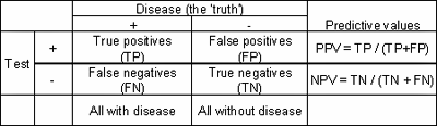
If we take a method with the following performance characteristics:
Sensitivity = 90 %
Specificity = 45 %
and we test four different populations, all with 100,000 individuals but with different prevalence of disease, we get differences in predictive values.
TABLE II: Test used in populations with different prevalence
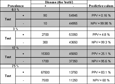
As the prevalence increases, so does the positive predictive value, while the negative predictive value decreases.
Hospitals may group patients into low-pretest-probability, medium-pretest-probability and high-pretest-probability patients. The pretest probabilities of DVT for these groups are often given as 3 %, 17 % and 75 % [8].
However, at one hospital the true prevalence of DVT in the low-pretest-probability group may be 7 %, at another hospital it may be 1 %. This means that the calculated NPV at one site would be several percentage points lower than at the other site due solely to the differences in prevalence (all other factors being equal).
Spectrum of disease/mix of VTE according to position
Ideally, the ability of a test to correctly identify patients with and without a specific disease should not vary between patients. However, the spectrum of disease found among patients in the population upon which the test is to be used will often have an influence on the clinical performance of the test.The spectrum of disease for VTE patients is primarily determined by the site of the DVT [10], distal (calf veins) or proximal (thigh veins) and whether PE is present or not.
A DVT in the calf veins is usually smaller and gives rise to lower D-dimer concentrations than a DVT in the thigh veins [10]. For this reason the risk of getting a false negative result is larger when the DVT is distal compared to the risk when the DVT is proximal.
Most D-dimer studies are performed in hospitals on patient populations with a predominance of proximal DVT. In an outpatient study including 393 patients with clinically suspected symptomatic DVT of the lower extremities [10] a total of 137 of 393 patients had a proven DVT and the majority (59 %) presented with distal DVT.
Twenty-eight out of 81 patients with distal DVT had a normal D-dimer, compared with two of 56 patients with proximal DVT. The sensitivity for distal DVT was only 65 % compared with 96 % for proximal DVT; the negative predictive values were 84 % and 99 %, respectively.
PE is usually associated with a higher D-dimer concentration than a DVT without PE [11]. This means that clinical sensitivities, specificities and predictive values cannot be compared between studies focusing on PE and DVT, respectively.
Assay performance – clinical sensitivity and specificity
A comparison of either clinical sensitivities or specificities without the other parameter is an arbitrary one, since changing the cut-off can increase sensitivities or specificities at the expense of lowering the other parameter [9]. A joint comparison of clinical sensitivity/specificity pairs may still be meaningful if the sensitivity and specificity of one method are both higher than the sensitivity and specificity of the other method.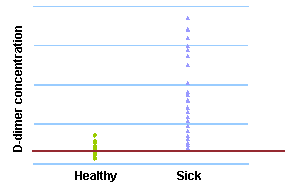
FIG. 1: Cut-off = 400 µg/L
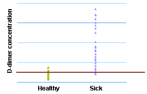
FIG. 2: Cut-off = 500 µg/L
The figures above are examples of the trade-off between clinical sensitivity and specificity. When 400 µg/L is chosen as cut-off, the sensitivity is 100 % and the specificity is 54 %. When the cut-off is increased to 500 µg/L, the sensitivity decreases to 92 % and the specificity increases to 79 %.
Gold standard used to establish diagnosis
The gold standard used to establish a VTE diagnosis is usually based on either ultrasound or angiography/venography.Ultrasound has a limited ability to image the calf veins where the thrombi are smaller [2]. This means that there is a risk of classifying patients with calf DVT as non-sick. This will give bias such as falsely increased sensitivity and negative predictive value because a negative D-dimer in combination with a false negative ultrasound DVT test will be registered (in error) as a true negative test.
A review of 26 studies [2] shows up to 9.2 % differences in NPV for each D-dimer test when evaluated versus a diagnostic test detecting all clots, and when evaluated versus a test detecting primarily the clots in the thigh.
It is also important to notice if all test persons were examined with a confirmatory method. In some studies only patients with a positive D-dimer test are examined with a confirmatory method. VTE is often asymptomatic, misdiagnosed, and unrecognized at death [12].
Some estimates of the likelihood of diagnosing DVT are as low as 10 %. Due to a lack of routine post-mortem examinations it is difficult to estimate the true incidence of VTE. We can only say that it is underestimated.
The fact that VTE is underestimated is also supported by a meta-analysis finding that ~70 % of the major (either fatal or contributing to death) pulmonary embolic events diagnosed at autopsy had been missed by the attending clinicians [13].
Falsely increased sensitivity and negative predictive value may be seen when a person has a negative D-dimer and a DVT that is not detected due to lack of examination.
Comparison with the same samples measured by both/all the assays to be compared
The ideal D-dimer method comparison is based on results obtained with both/all assays on the same samples and when all tests persons are examined with a confirmatory method.Less ideal is a comparison based on results obtained with both/all assays on the same samples where only all the test persons with discrepant results (below cut-off with one assay, above cut-off with the other assay) are examined with a confirmatory method.
It should be kept in mind that a comparison based on results obtained with both/all assays on the same samples can be directly used to compare the relative performances of the assays, but the relative performances may be different if the assays are tested on another population with a different prevalence, a different spectrum of disease, etc.
References1. Heim SW, Schectman JM, Siadaty MS et al. D-dimer testing for deep venous thrombosis: a metaanalysis. Clin Chem 2004; 50(7): 1136-47
2. Philbrick JT, Heim S. The D-dimer test for deep venous thrombosis: gold standards and bias in negative predictive value. Clin Chem 2003; 49(4): 570-74
3. Wells PS, Anderson DR, Rodger M et al. Evaluation of D-dimer in the diagnosis of suspected deep-vein thrombosis. N Engl J Med 2003; 349: 1227-35
4. Dempfle CE. Validation, calibration, and specificity of quantitative D-dimer assays. Semin Vasc Med 2005; 5(4): 315-20
5. Brotman DJ, Segal JB, Jani JT et al. Limitations of D-dimer testing in unselected inpatients with suspected venous thromboembolism. Am J Med 2003; 114: 276-82
6. Dempfle CE: D-dimer testing and venous thromboembolism: four view points. J Thromb Haemost 2005; 3: 377-79
7. Harper PL, Theakston E, Ahmed J et al. D-dimer concentration increases with age reducing the clinical value of the D-dimer assay in the elderly. Intern Med J 2007; 37(9): 607-13
8. Wells PS, Anderson DR, Bormannis J et al. Value of assessment of pretest probability of deep-vein thrombosis in clinical management. Lancet 1997; 350: 1795-98
9. CLSI/NCCLS document EP12-A2 User Protocol for Evaluation of Qualitative Test Performance; Approved Guideline 2nd Edition. Vol. 28 No. 3. 2008
10. Jennersjö CM et al. Normal D-dimer concentration is a common finding in symptomatic outpatients with distal deep vein thrombosis. Blood Coagul Fibrinolysis 2005; 16: 517-23
11. Tick LW et al. High D-dimer levels increase the likelihood of pulmonary embolism. J Intern Med 2008; 264(2): 195-200
12. Cohen AT et al. Venous thromboembolism (VTE) in Europe – the number of VTE events and associated morbidity and mortality. Thromb Haemost 2007; 98(4): 756-64
13. Task Force Report of the European Society of Cardiology. Guidelines on diagnosis and management of acute pulmonary embolism. Eur Heart J 2000; 21: 1301-36


Register for the related webinar
The Benefit of Using a D-dimer Assay with a High Clinical Specificity
Presented by Karin Strandberg, MD, PhD, Assoc. Prof.
Join the webinar
Acute care testing handbook
Get the acute care testing handbook
Your practical guide to critical parameters in acute care testing.
Download now
Scientific webinars
Check out the list of webinars
Radiometer and acutecaretesting.org present free educational webinars on topics surrounding acute care testing presented by international experts.
Go to webinars