Printed from acutecaretesting.org
April 2013
Why measure blood gases? A three-part introduction for the novice. Part 3.


Reference (normal) range for blood gas parameters under discussion here:
pO2(a) 10.6-13.3 kPa (80-100 mmHg)
sO2(a) 95-98 %
Reference (normal) range for O2 saturation by pulse oximetry (SpO2) 95-98 %
HYPOXEMIA, ISCHEMIA AND TISSUE HYPOXIA
The principal clinical value of measuring pO2(a) and sO2(a) is to detect hypoxemia, which can be defined as a reduced amount of oxygen in blood. Hypoxemia is diagnosed if pO2(a) and/or sO2(a) are below the lower limit of their respective reference range. However, as will hopefully be made clear, normal pO2(a) and/or sO2(a) do not necessarily exclude a diagnosis of hypoxemia.
Although hypoxemia can be indicative of a range of respiratory and non-respiratory diseases, it does not of itself have major significance for health. Blood is merely the means for transporting oxygen to tissue cells and hypoxemia is only really significant because it threatens adequate oxygenation of tissue cells.
Hypoxia, which compared with hypoxemia is the more significant state, is defined as insufficient oxygen supply to cells for normal aerobic metabolism to be sustained. Hypoxia results in cell death, organ failure and, if not corrected, is ultimately fatal. The significance of hypoxemia then is that it can, if sufficiently severe, result in hypoxia.
But hypoxemia is not the only cause of hypoxia; the other major cause is reduced blood flow (ischemia). Myocardial infarction provides a striking example of potentially fatal local tissue hypoxia being caused not by hypoxemia, but by ischemia, due to thrombosis of a coronary artery.
Mild hypoxemia (defined below) does not usually result in hypoxia but in patients with cardiac insufficiency the combined effect of mild hypoxemia and reduced blood flow threatens tissue oxygenation to a greater extent than either hypoxemia or reduction of cardiac output would alone.
By the measurement of pO2(a) and sO2(a), arterial blood gases thus provide vital but incomplete information for full assessment of patient oxygenation status.
RELATIONSHIP BETWEEN pO2(a) AND sO2(a)
Although already discussed in the first article of this series, it is useful to remind here that pO2(a) and sO2(a) are different, but related measures of arterial blood oxygenation. pO2(a) is a measure of the pressure exerted by the very small fraction (1-2 %) of total oxygen in arterial blood that is dissolved in blood plasma, whereas sO2(a) reflects the remaining 98-99 % of total oxygen in arterial blood that is bound to hemoglobin in red blood cells. To be specific, sO2(a) is the percentage of hemoglobin in arterial blood that is capable of binding oxygen, and is saturated with oxygen:
sO2(a) = O2Hb / (O2Hb + HHb) × 100
(where O2Hb = concentration of oxygenated hemoglobin in arterial blood and HHb = concentration of deoxygenated hemoglobin in arterial blood).
sO2(a) obviously cannot possibly be greater than 100 %. pO2(a) is the major determinant of sO2(a), and the relationship between the two parameters is described by the oxygen dissociation curve, ODC (see article 1). From this graph it is evident that there is a positive correlation between pO2(a) and sO2(a); as pO2(a) rises, so too does sO2(a). It is also evident from the sigmoidal shape of the curve that the positive correlation is non-linear.
This means that despite increasing pO2(a) above around 8.0 kPa, there is very little increase in sO2(a). So, although pO2(a) reflects only a tiny fraction of the oxygen in blood, it is a much more sensitive measure of blood oxygenation than sO2(a) if blood oxygenation is increased, normal or only slightly reduced (cf. the flat part of the ODC).
Below are some pO2(a) and corresponding sO2(a) values that characterize normal blood oxygen (normoxemia), reduced blood oxygen (hypoxemia) and increased blood oxygen (hyperoxemia). The sO2(a) values assume normal acid-base balance (they are lower in acidosis and higher in alkalosis)
| Normoxemia | pO2(a) 10.6 kPa (80 mmHg) | sO2(a) 96 % | |
| pO2(a) 13.3 kPa (100 mmHg) | sO2(a) 98 % | ||
| Hypoxemia (mild) | pO2(a) 9.3 kPa (70 mmHg) | sO2(a) 94 % | |
| Hypoxemia (moderate) | pO2(a) 8.0 kPa (60 mmHg) | sO2(a) 91 % | |
| Hypoxemia (severe) | pO2(a) 6.0 kPa (45 mmHg) | sO2(a) 80 % | |
| Hyperoxemia | pO2(a) 16.0 kPa (120 mmHg) | sO2(a) 98 % | |
| Hyperoxemia (marked) | pO2(a) 20.0kPa (150 mmHg) | sO2(a) 99 % |
It is noteworthy that as pO2(a) rises from 9.3 kPa (mild hypoxemia) through 12.0 kPa (normoxemia) and onwards to 16.0 kPa (hyperoxemia), sO2(a) only rises from 94 % to 98 %. Thus, perhaps counterintuitively, no great increase in the total amount of oxygen in arterial blood is achieved by raising pO2(a) from 9.3 kPa to 16.0 kPa. By contrast, reduction of pO2(a) by only 3.3 kPa from 9.3 kPa to 6.0 kPa is associated with significant reduction in sO2(a) from 94 % to 80 %, and therefore significant reduction in the total amount of oxygen present in blood. Remember, it is the oxygen bound to hemoglobin that accounts for nearly all (>98 %) of the oxygen in blood.
Respiratory failure is defined by pO2(a) less than 8 kPa (sO2(a) less than 91 %). Below the degree of hypoxemia that these levels represent, there is increasing risk of hypoxia, even if cardiac output is not compromised. This degree of hypoxemia would usually trigger prescription of supplemental oxygen therapy to ensure adequate tissue oxygenation.
NORMAL pO2(a) AND sO2(a) DOES NOT NECESSARILY MEAN THAT BLOOD CONTAINS SUFFICIENT OR EVEN NORMAL AMOUNT OF OXYGEN
Although it is true that for normal blood oxygenation pO2(a) and sO2(a) must both be normal, the finding of pO2(a) and/or sO2(a) within their normal range is not sufficient evidence, of itself, to be assured of normal or even adequate blood oxygenation. This is because there is a third element to be considered: the amount (or concentration) of hemoglobin.
There must be sufficient hemoglobin to carry the oxygen transferred from inspired air to blood during pulmonary gas exchange. sO2(a) is a measure of the percentage of hemoglobin that is capable of binding oxygen and is saturated with oxygen. Thus sO2(a) can remain within the reference range even if there is insufficient hemoglobin, and pO2(a), of course, is unaffected by hemoglobin concentration.
Anemia (defined by reduced total hemoglobin concentration) therefore represents a relatively common condition in which, despite normal pO2(a) and sO2(a), blood oxygenation is reduced. Consideration of hemoglobin concentration is as vital as consideration of pO2(a) and sO2(a) when assessing patient blood oxygenation status.
Adequate blood oxygenation depends not only on a sufficient amount of hemoglobin but also on the capability of that hemoglobin to bind reversibly with oxygen; it must be functional. There are several pathological situations in which hemoglobin is present in normal amount but a significant percentage is dysfunctional and incapable of binding oxygen, and in this circumstance pO2(a) is normal (because it is a measure of oxygen dissolved in blood plasma, not of that bound to hemoglobin) but sO2(a) is reduced.
Carbon monoxide poisoning, which results in increased concentration of the dysfunctional hemoglobin species, carboxyhemoglobin (COHb), is the most common of these pathological situations. It is rare for there to be a total disconnect between pO2(a) and sO2(a) values, but carbon monoxide poisoning is representative of conditions (collectively called the dyshemoglobinemias) in which this is usually the case.
This raises an important issue regarding the way sO2(a) values are generated during blood gas analysis. Most modern blood gas analyzers have an incorporated CO-oximeter that allows direct (spectrophotometric) measurement of sO2(a). In the absence of a CO-oximeter, sO2(a) generated during blood gas analysis is calculated from measured pO2(a); the calculation is based on the oxygen dissociation curve.
In nearly all circumstances calculated sO2(a) (generated by some blood gas analyzers) is a clinically acceptable approximation of the true (measured) sO2(a) value, but in the context of excessive amount of non-functional hemoglobin (dyshemoglobin), for example in carbon monoxide poisoning, that is not the case. For accurate assessment of blood oxygenation among patients suffering carbon monoxide poisoning and methemoglobinemia it is important that a blood gas analyzer with incorporated CO-oximeter be used.
CAUSES OF HYPOXEMIA (REDUCED pO2(a), sO2(a))
There are five mechanisms that give rise to hypoxemia:
- Partial pressure of oxygen of inspired air reduced (altitude effect)
- Global alveolar hypoventilation (common)
- Mismatch of alveolar ventilation/perfusion (the most common)
- Cardiovascular shunting (rare)
- Diffusion limitation (usually only a contributory mechanism)
1. Partial pressure of inspired air (pO2(I)) reduced
In health pO2(a) is determined by the partial pressure of oxygen in alveolar air (pO2(A)), which in turn is determined by the partial pressure of oxygen in inspired air (pO2(I)). Reduction in pO2(I) (essentially breathing in less oxygen) inevitably results in reduced pO2(A) and therefore reduced pO2(a)/sO2(a). pO2(I) is dependent on the pressure of atmospheric air (barometric pressure).
At sea level, barometric pressure is 760 mmHg (101 kPa) so since air comprises 21 % oxygen, pO2(I) at sea level is 21 kPa (21 × 101 / 100). (In fact it is 1.4 kPa less than this due to the effect of water vapor that humidifies inspired air and effectively dilutes the oxygen). Barometric pressure decreases with altitude, so that for example, at 3000 meters above sea level where barometric pressure is 72 kPa, pO2(I) is just 13.7 kPa (21 × 72 / 100 – 1.4) and resulting pO2(a) is around 7.0 kPa, sO2(a) 85 % (assuming no compensatory increase in depth and rate of respiration). In short, hypoxemia can be caused by breathing air at high altitude. It is important to note that published reference (normal) ranges for pO2(a) and sO2(a) assume breathing ambient air (21 % oxygen) at sea level.
2. Global alveolar hypoventilation
Alveolar ventilation is the volume of air breathed in per minute that reaches the alveoli and takes part in gas exchange with blood across the alveolar/capillary membrane. Global alveolar hypoventilation means that less oxygen than normal is available in all alveoli for diffusion to blood (i.e. pO2(A) is reduced) and pO2(a) inevitably falls. Since, as explained in the first article in this series, ventilation is regulated via pCO2(a)-sensitive chemoreceptors to maintain pCO2(a) within normal limits, alveolar hypoventilation is necessarily associated with increased pCO2(a) and consequent respiratory acidosis (discussed in the second article).
If pCO2(a) is within normal limits or reduced, then global alveolar hypoventilation cannot be the explanation for hypoxemia. In global alveolar hypoventilation every 1 mmHg (0.133 kPa) increase in pCO2(a) is accompanied by 1 mmHg (0.133 kPa) decrease in pO2(a).
There are a number of diseases/conditions that can cause global alveolar hypoventilation and consequent hypoxemia (some are listed in Table I). The problem commonly lies outside of the lungs when it reflects disturbance of neural or muscular action required for regulating the rate and/or mechanics of breathing.
As might be expected the list includes many conditions that also appear on the list of causes of respiratory acidosis (article 2). [Respiratory failure (essentially failure of pulmonary gas exchange) is diagnosed if pO2(a) is less than 8.0 kPa. If pCO2(a) is normal or reduced, the condition is called Type 1 respiratory failure; if pCO2(a) is raised, the condition is called Type 2 respiratory failure. It should be evident from the discussion above that alveolar hypoventilation is a condition that can, if sufficiently severe, give rise to Type 2 respiratory failure, but never to Type 1 respiratory failure].
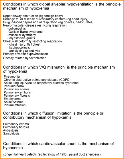
Table I: Causes of hypoxemia (reduced pCO2(a), sO2)
3. Ventilation-perfusion mismatch
This is the most common of the five mechanisms that give rise to hypoxemia. Optimum pulmonary gas exchange and therefore optimum blood oxygenation depend on equality between ventilation and blood perfusion of all alveoli. No matter how well ventilated a particular alveolus is, gas exchange at that alveolus cannot occur if it is not perfused with blood.
Likewise, despite optimum blood flow through the capillaries of an alveolus, gas exchange will not occur if the alveolus does not contain inspired air, i.e. is not ventilated. For optimum pulmonary gas exchange, ventilation rate (V) must match blood flow rate (Q), that is to say, the ratio V/Q must ideally be 1.
Ventilation-perfusion mismatch refers to conditions in which hypoxemia occurs because due to disease there are areas of lung in which V/Q is significantly greater or less than 1. As an example, consider the patient with pneumonia whose lungs contain areas that are consolidated by pus.
The V/Q ratio in these disease-affected areas is reduced due to reduced ventilation in the face of perfusion that remains unaffected by the disease process. Pulmonary embolism, on the other hand, exemplifies those conditions in which hypoxemia results from a raised V/Q ratio (i.e. area of lung in which alveoli are optimally ventilated but inadequately perfused). The term intrapulmonary right-to-left shunt is used to describe those areas of lung where V/Q is actually zero (adequate perfusion but no ventilation).
A significant feature of hypoxemia due to V/Q mismatch is that pCO2(a) usually remains, counterintuitively, normal. This is in large part because the hyperventilation that hypoxemia induces increases excretion of CO2 from unaffected alveoli and this compensates for the reduced excretion of CO2 that necessarily occurs across disease-affected (poorly ventilated) alveoli.
Hyperventilation of healthy alveoli cannot compensate for hypoxemia due to V/Q mismatch in this way because there is no capacity to increase the oxygen in capillary blood serving healthy alveoli – it is already maximally saturated with oxygen. A rapidly rising pCO2(a) in such cases may be a poor prognostic sign because it can signal exhaustion from the effort of overbreathing, and possible imminent respiratory collapse (cessation of ventilation).
V/Q mismatch is thus usually associated with Type 1 respiratory failure (pO2(a) < 8.0 kPa, normal pCO2(a) but can also be associated with Type 2 respiratory failure (pO2(a) < 8.0 kPa , increased pCO2(a)) if compensatory increase in ventilation is inadequate.
The hypoxemia associated with many common pulmonary and non-pulmonary diseases is usually the result of V/Q mismatch (Table I).
4. Cardiovascular shunt
Hypoxemia can result from structural congenital heart defects that result in abnormal shunting of blood through a circulation that does not allow a significant proportion of blood access to the pulmonary circulation and therefore to ventilated alveoli. This is called a right-to-left shunt, meaning that effectively deoxygenated blood leaks from the right heart to the left heart. Tetralogy of Fallot is the most common congenital heart defect to cause right-to-left shunt-induced hypoxemia.
Defects that result in left-to-right shunt (e.g. patent ductus arteriosus) can also be associated with hypoxemia although the mechanism of the hypoxemia in these cases is a little more opaque than in the case of right-to-left shunts. In left-to-right cardiac shunts the problem is increased blood flow (Q) through alveolar capillaries (consequent on the extra volume of leaked blood to the pulmonary circulation), and resulting reduced V/Q ratio.
5. Diffusion limitation
Oxygenation of blood depends on oxygen diffusing from alveoli to blood due to the concentration gradient that exists between alveoli and mixed venous blood flowing through alveolar capillaries (pO2(A) greater than pO2(v)). Oxygen must diffuse across the alveolar/capillary membrane (sometimes referred to as the blood-gas barrier). Effective rate of diffusion is made possible by the extreme thinness of this barrier (of the order 0.2-0.3 mm). A number of lung diseases result in thickening of the barrier and resulting lengthening of the diffusion path; this inevitably reduces the rate of diffusion.
The pO2 of mixed venous blood flowing past a disease-thickened barrier will not reach equilibrium with the pO2 of alveolar air, in which case hypoxemia may occur. The tendency to hypoxemia due to diffusion limitation is enhanced during exercise because this increases blood flow, thereby reducing the time that blood is exposed to alveoli and the oxygen they contain. It is unusual for diffusion limitation to be the sole mechanism responsible for hypoxemia but it is a contributory factor in a number of lung diseases (Table I).
SUPPLEMENTAL OXGEN THERAPY – THE ONLY MAJOR CAUSE OF HYPEROXEMIA (INCREASED pO2(a), sO2(a))
Supplemental oxygen is therapeutically effective for correction of hypoxemia because it increases the fraction of inspired oxygen (FO2(I)) and thereby the partial pressure of inspired air (pO2(I)). Increased pO2(I) in turn results in increased partial pressure of alveolar air (pO2(A)) and ultimately increased partial pressure of arterial blood (pO2(a)). FO2(I) is usually expressed on a scale of 0 (0 %) to 1 (100 %) so the FO2(I) of the air we breathe is 0.21 (21 %) and resulting pO2(I) is 20.0kPa (at sea level). Depending on the mode of oxygen delivery, FO2(I) can range from 0.24 to 1.00 (24-100 %).
Oxygen therapy (i.e. increasing FO2(I)) is effective in correcting hypoxemia due to global hypoventilation and V/Q mismatch but is less effective among patients who have significant right-to-left shunts (either intrapulmonary or cardiovascular) because in these cases blood is bypassing alveoli and no matter how high the pO2(A), pO2(a) of shunted blood will remain unaffected.
Knowledge of the FO2(I) is important for interpretation of pO2(a)/ sO2(a) results among patients receiving supplemental oxygen. The important questions must be:
- is the pO2(a) appropriately high for FO2(I)?
- does the pO2(a)/sO2(a) indicate adequate oxygenation of blood (i.e. is the FO2(I) sufficiently high)?
A useful rule of thumb for testing the appropriateness of pO2(a) is that the difference between FO2(I) (expressed as a percentage) and pO2(a) (kPa) should not exceed 10. So that, for example, an individual receiving 30 % oxygen should by this rule of thumb, if oxygenation is unimpaired, have a pO2(a) > 20.0 kPa. The finding of pO2(a) within the reference range (10.6-13.3 kPa) is clearly inappropriately low for an individual receiving 30 % oxygen, indicating pathological impairment of oxygenation. Such a result, however, does indicate that oxygen therapy is compensating for the impairment and that FO2(I) of 0.30 (30 %) is sufficient to ensure more than adequate blood oxygenation for this patient.
sO2(a) versus SpO2 – A NOTE ON PULSE OXIMETRY
Measurement of pO2(a) and sO2(a) during arterial blood gas analysis (ABG) is the gold-standard method for assessment of patient blood oxygenation status. Pulse oximetry is an alternative less accurate and less precise technique that despite these and other limitations has become ubiquitous throughout clinical medicine. It has the great advantage over ABG of being non-invasive (does not require blood sampling) and enables safe and easy continuous monitoring of blood oxygenation status via a small probe that is simply clipped on the fingertip, earlobe or toe.
A pulse oximeter provides an estimation of (not a direct measurement of) arterial oxygen saturation (sO2(a)) and this estimation is based on the differences in the absorbance of light at two wavelengths (~660 nm and ~940 nm) of oxyhemoglobin, O2Hb (i.e. oxygenated hemoglobin) and deoxyhemoglobin, HHb (i.e. deoxygenated hemoglobin). The pulse oximeter probe comprises two elements – a light emitter and a light detector – that are sited opposite each other on either side of the fingertip. The light emitter ”shines” light of two wavelengths (660 nm and 940 nm) at the fingertip and the detector detects the amount of light at the two wavelengths on the other side of the fingertip. Crucially, the amount of light at the two wavelengths absorbed by the fingertip is predictably related to the concentrations of O2Hb and HHb in blood”pulsing” through the capillary bed of the fingertip. Calculation of O2Hb and HHb concentration from absorbances at the two wavelengths allows, in turn, calculation of oxygen saturation:
Oxygen saturation (%) = [O2Hb / O2Hb + HHb (i.e. total Hb)] × 100
The oxygen saturation parameter generated by pulse oximetry is SpO2 and the justification for using pulse oximetry to assess blood oxygenation status depends on the now well-validated notion that SpO2 is a reliable and clinically acceptable estimate of sO2(a) in nearly all clinical circumstance; SpO2 is usually within 1-3 % of sO2(a).
It must be emphasized that in generating SpO2, a pulse oximeter only provides an estimation of arterial oxygen saturation (sO2(a)). It simply cannot measure pO2(a), pCO2(a) or pH, so unlike ABG it only provides a partial assessment of pulmonary gas exchange and provides no information about patient ventilatory and acid-base status.
It is important to know the limitations of pulse oximetry and appreciate the clinical conditions in which SpO2 might be a less than reliable estimate of sO2(a). Accuracy and precision of pulse oximetry is reduced if oxygen saturation is markedly reduced and so is increasingly less reliable if sO2(a) falls below around 80 %.
For optimum performance pulse oximetry requires normal pulsatile arterial blood flow, and reduced perfusion of the measuring site – as might occur in the context of hypovolemia, severe hypotension, clinical shock etc. – results in falsely low SpO2. Irregular heart rhythms can reduce the reliability of SpO2 readings, as can untoward motion of the pulse oximeter probe that occurs, for example, in the shivering patient.
This all means that pulse oximetry is generally less reliable in assessing oxygenation status among the critically ill than among other patient groups. Improvement in pulse oximeter design continues, and new-generation pulse oximeters are less susceptible to some of the limitations described above.
One very specific limitation that persists relates to the fact that the measuring system in pulse oximeters is unable to distinguish functional hemoglobin from dysfunctional hemoglobins (carboxyhemoglobin, COHb and methemoglobin, MetHb). This means that SpO2 is not an accurate estimation of sO2(a) in patients suffering conditions in which COHb or MetHb levels are abnormally high.
For example, in patients with carbon monoxide poisoning, which is characterized by increased COHb, measured (but not calculated) sO2(a) is, as discussed above, reduced, indicating often severe hypoxemia. The SpO2 in such cases remains normal (or even raised to 99 %), providing comforting but dangerously misleading evidence of adequate oxygenation. Pulse oximetry should not be used to monitor oxygenation status in patients with suspected or confirmed carbon monoxide poisoning or in patients with suspected or confirmed methemoglobinemia. In such cases arterial blood gas (with CO-oximetry) is obligatory for accurate assessment of oxygenation status.
A marked disconnect between SpO2 and sO2(a) (SpO2 > sO2(a)) is also evident if venous rather than arterial blood has been inadvertently sampled (an error that occasionally occurs in clinical practice). Under such circumstance the ”sO2(a)”, which is actually sO2(v-), provides erroneous evidence of severe hypoxemia (normal sO2(v-) around 75 %), but the SpO2 (unaffected by the sampling error) reflects true arterial oxygenation. In this way pulse oximetry provides a useful quality safety check of arterial blood gases in that it can alert to this rare, but clinically misleading, sampling error.
References+ View more
- West J. Respiratory physiology - the essentials. 8th ed. Baltimore: Lippincott Williams & Wilkins, 2008.
- Dominiczak M, Szczepanska-Konkel M. Regulation of hydrogen ion concentration (acid-base balance) In: Medical Biochemistry. 3rd ed. Mosby, 2009.
- Waugh A, Grant A. The respiratory system. In: Ross and Wilson anatomy and physiology in health and illness. 11th ed. Churchill-Livingstone, 2010.
- Hennesey I, Japp A. Arterial blood gases made easy. Churchill-Livingstone, 2007.
References
- West J. Respiratory physiology - the essentials. 8th ed. Baltimore: Lippincott Williams & Wilkins, 2008.
- Dominiczak M, Szczepanska-Konkel M. Regulation of hydrogen ion concentration (acid-base balance) In: Medical Biochemistry. 3rd ed. Mosby, 2009.
- Waugh A, Grant A. The respiratory system. In: Ross and Wilson anatomy and physiology in health and illness. 11th ed. Churchill-Livingstone, 2010.
- Hennesey I, Japp A. Arterial blood gases made easy. Churchill-Livingstone, 2007.
May contain information that is not supported by performance and intended use claims of Radiometer's products. See also Legal info.


Acute care testing handbook
Get the acute care testing handbook
Your practical guide to critical parameters in acute care testing.
Download now
Related webinar
Evolution of blood gas testing Part 1
Presented by Ellis Jacobs, PhD, Assoc. Professor of Pathology, NYU School of Medicine.
Watch the webinar